A summary of the chapter “The Dynamics of Breathing” from the book Yoga Anatomy [1]
The book “Yoga Anatomy” by Leslie Kaminoff is very clearly organized. Dynamics of Breathing : In the introductory part there is general knowledge about breathing, spine, skeleton and muscles. The main part explains the most important asanas and their effect on organs, muscle groups and joints. Furthermore, the Sanskrit terms of the Asanas are translated.
- What actually happens in the body during different yoga exercises, breathing, joint and muscle actions?
- Which muscles and joints are involved in individual yoga poses?
- Which muscles are active, which are stretched? -When to use which breathing techniques?
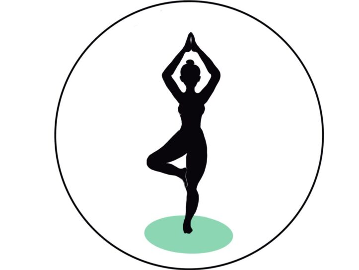
Asanas & Breathing an Inseparable Combination
In their book, authors Kamminoff and Matthews describe in great detail the dynamics of breathing as well as the role of the diaphragm. Breathing is viewed holistically in the context of yoga exercises.
Dynamics of breathing
The dynamics of respiration is derived from the functional principle of a cell. Nutrients pass through the cell membrane and are metabolized internally to provide energy. During the metabolic process waste products are produced, which have to leave the membrane again. These cellular functions are called prana (inhalation) and apana (exhalation). The structural properties of the membrane (permeability, stability) are described by the terms sthira (firm, compact, strong) and sukha (light, gentle, mild).
Breathing is the movement of air. This air enters the lungs from the environment and is released from the lungs back into the environment. Oxygen metabolism then takes place. Oxygen and carbon dioxide is exchanged between the lungs and the blood, and between the blood and the cells of the body.
The path of respiration
Several organs and muscles are involved in this chemical and biomechanical process. The pathway begins with the nose, which breathes in air enriched with oxygen. Through the throat, the air enters the larynx, and is passed to the lungs. The capillaries in the lungs transfer the oxygen to the blood, which uses it to supply the body’s cells. Resulting carbon dioxide (CO2) is then transported by the blood back to the lungs and finally returns to the environment through the larynx, pharynx and nose.
The most important muscle in breathing is the diaphragm
The diaphragm begins between the third and fourth ribs and is attached to the heart. Moving upward, the diaphragm becomes a tendon plate. It is stretched round like a parachute. Because on the left side the heart is pressed down and on the right side the liver is pressed up. The parachute extends in permanent asymmetry to the second or third lumbar vertebra and is attached there to four points, namely the sternum, ribs, costal arches and loins.
Abdominal breathing
The diaphragm separates the chest cavity, including the lungs and heart, from the abdominal cavity, where the internal organs are located. The internal organs such as the stomach, intestines, spleen, liver as well as kidneys are not directly involved in the mechanics of breathing because they are naturally supplied with oxygen. When the lungs fill with fresh oxygenated air during inhalation, their volume increases and the lungs expand in all directions. The diaphragm, as well as the intercostal muscles located above the diaphragm, then push the organs away from above to allow the lungs to expand. Since the abdominal organs have no internal space to escape, they expand visibly from the outside and the abdominal wall lifts. On exhalation, the diaphragm lifts again supported by the oblique abdominal muscles and the transversus abdominis muscle, forcing air out of the lungs.
Chest breathing
The respiratory accessory muscles of the intercostals and the oblique abdominal muscles merge, which means that only one of them is active at any given time, while the other muscle is relaxed. Deep breathing with a tense abdomen is difficult for this reason. Tightening the lower abdominal muscles or even an above-average filled gastrointestinal tract can push the lungs more to the sides and upward. This breathing is called chest breathing.
Abdominal and thoracic breathing engage muscle groups at different intensities. One way in which forces are redistributed is by closure of the larynx. This closure can occur through two of four possible positions in the vocal cords. When awake or asleep, the vocal cords are open to different degrees. Therefore, air is transported up and down through the muscles of the intercostals, the abdominal muscles mentioned above, and the diaphragm.
Ujjayi breathing
In Ujjayi breathing, one nostril is closed. Thus, the breaths are slowed down. Automatically, the breathing is much more conscious and deepened. In this breathing, the flow of breath is both perceptible and audible, like the sound of the sea. As a result, the activity of the muscles is fully carried out and the spine stretches into an upright position. The complete closure of the larynx is called Jalandara Bandha in Sanskrit. In order to close the vocal cords completely and to make it easier to hold the breath, the chin is also drawn to the sternum here.
The term pranayama is divided into prana (the life) and yama (control). Controlling our breath can help us find the intrinsic GG.
The book “Yoga Anatomy” is a good reference book to prepare the yoga class in depth and to address students’ concerns about muscle tension in the shoulder, neck and lower back.
Sources:
[1] Leslie Kaminoff: Yoga Anatomie, riva Verlag, 2018, 4. Auflage